
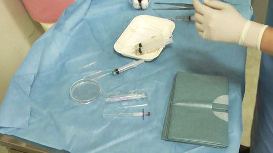
Preparations for RI examination
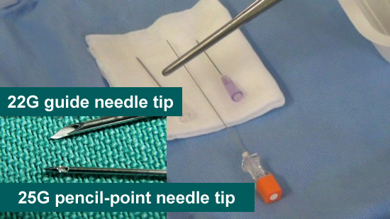
●Puncture needle
25-gauge pencil-point needle (Puncture needle)
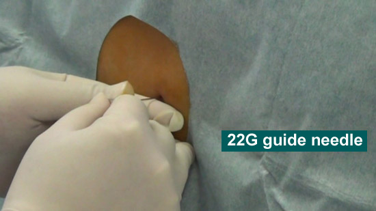
22-gauge needle (Guide needle)
●Extension tube

●Local anesthesia
24-gauge needle (Local anesthesia needle)
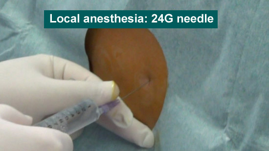
Local anesthetic
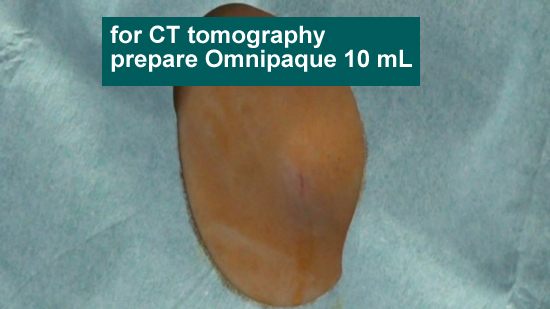
●Contrast medium
Omnipaque for computed tomography (CT) 10 mL
Indium 1 mL
●Measurement rod
Description
(1)Local anesthesia (24-gauge needle)
(2)Prepare Omnipaque 10 mL for a syringe beforehand
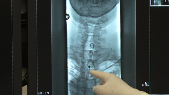
(3)Insert a guide needle (22-gauge needle) just in front of the dura mater
(4)Insert a pencil-point puncture needle into the dura mater
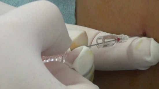
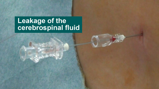
(5)Check the leakage of the cerebrospinal fluid
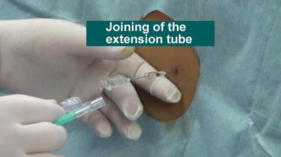
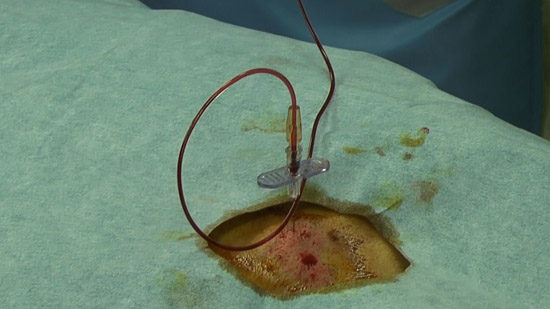
(6) Join an extension tube
Conduct the procedure with the extension tube hereafter
(7) Measurement of the lumbar puncture pressure
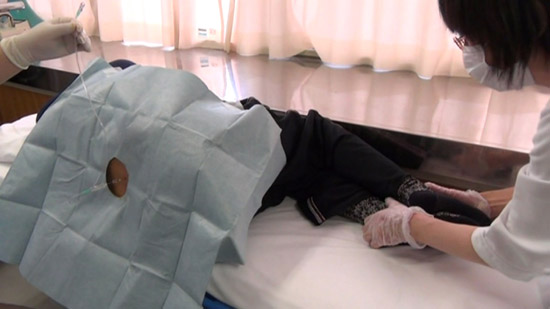
First, relax the posture of patients and lower the abdominal muscle pressure
Measure the pressure with a glass rod placed in parallel to the extension tube
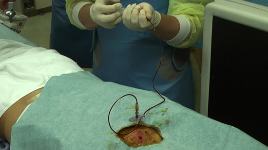
(8)Omnipaque 10 mL infusion
(9)Successively, inject indium 1 mL
Inject indium steadily using air pressure
(10)Remove all components from a guide needle

Blood patch
Perform blood patches at two regions for patients with idiopathic cerebrospinal fluid hypovolemia.
Supposing the leakage site is dorsal, conduct the puncture and blood patch at two regions on the dorsal and upper lumbar vertebrae of patients
Preparation
●Contrast medium
●Omnipaque (for myelography)
※ Differentiate an autologous blood syringe with a color needle
●Autologous blood syringe 20 cc - two
※Enter contrast medium 4 cc in it beforehand (about 20 cc)
●Local anesthesia (24-gauge needle)
●Puncture set
Syringe (glass) for epidural puncture
24-gauge puncture needle
●Saline
Description
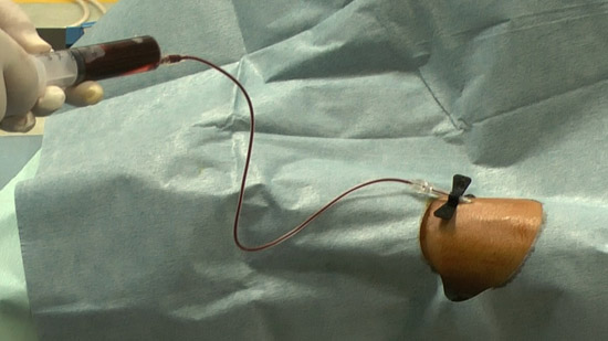
(1)Collect the blood of patients with autologous blood syringe
Because the contrast medium is administered beforehand, collect 16 cc blood twice
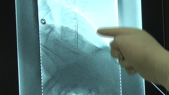
(2)Set the positioning by checking the puncture site along with the image on the radiological screen.
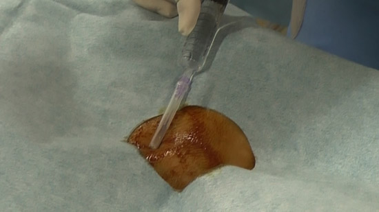
(3)Administer local anesthesia.
(4)Conduct an epidural puncture by the resistance loss method, checking with the image on the radioscopy screen.
※Inject the contrast medium and check it again. Capture a confirmation image at the same time.
(5)Connect an extension tube to the puncture needle and fill the autologous blood syringe with the collected blood (mixture contrast medium).
Check with the image on the radioscopy screen.
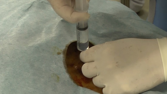
(6)Infuse the autologous blood.
While communicating with the patient, observe the patient’s condition.
Check subjective physical condition of patients after 10 cc infusion and capture a projectional image.
Successively, inject all the autologous blood.
Capture projectional images at the end of infusion.
Written by
Dr. Eiji Moriyama
Fukuyama Medical Center Hospital NeuroSurgery


Copyright (C) 2015 CSF JAPAN All Rights Reserved.